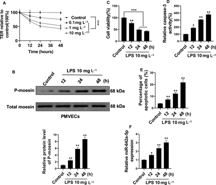
Fig. 1.

miR‐642a‐5p was induced by LPS stimulation in PMVECs. (A) Cultured human PMVECs grown to confluence on gold electrodes were treated with recombinant LPS (0.1, 1 or 10 mg·L−1), and changes in TER were measured at 12, 24 or 48 h. (B) Time‐dependent changes of phosphorylation of moesin in human PMVECs induced by 10 mg·L−1 LPS were detected by western blot. (C) The cell viability of human PMVECs induced with 10 mg·L−1 LPS for 12, 24 or 48 h was measured by MTT. (D, E) The apoptosis of human PMVECs treated with 10 mg·L−1 LPS for 12, 24 or 48 h was measured using the caspase‐3 activity assay and flow cytometry analysis. (F) The level of miR‐642a‐5p in human PMVECs treated with 10 mg·L−1 LPS for 12, 24 or 48 h was determined by qRT‐PCR. Two‐way ANOVA and a Bonferroni post hoc test (A), or one‐way ANOVA and a Dunn's post hoc test were used (B–F). Experiments were repeated independently in triplicate. Data were presented as mean ± SD. *P < 0.05, **P < 0.01, ***P < 0.001 versus control.
