
Figure 1.
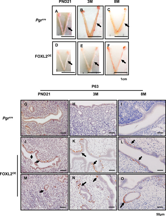
FOXL2OE mice displayed thin uterus and stratified uterine epithelium. The representative uterine images of Pgrcre (A–C) and FOXL2OE mice (D–F) at PND21 (A and D), 3 M (B and E), and 8 M (C and F). The uteri were much thinner in FOXL2OE mice compared with Pgrcre mice at 3 M and 6 M. Basal cell marker, P63, staining in the uterus of Pgrcre (G–I) and FOXL2OE mice (J–O) at PND21 (G, J, and M), 3 M (H, K, and N), and 6 M (I, L, and O). P63 positive basal cells in the uterus indicate epithelium stratification. They were found at the basal side of some luminal epithelium (J–L) and some glandular cells (M–O) in the FOXL2oe uterus. PND: postnatal day; M: months old. Arrow indicates stratified epithelium. N = 3 for each genotype and age group. Arrow in A–F refers one uterine horn. Arrow in J–O indicates stratified epithelium.