Figure 3.

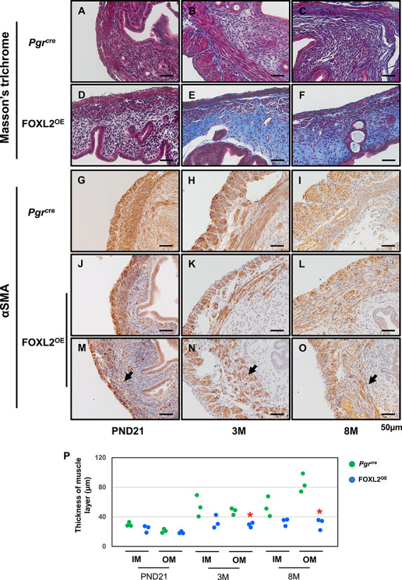
Stroma fibrosis and defective myometrium in FOXL2OE mice. Masson’s trichrome staining in Pgrcre (A–C) and FOXL2oe (D–F) uterus at PND21 (A and D), 3 M (B and E), 8 M (C and F). Increased blue staining in the stroma of FOXL2OE uterus suggested increased collagen deposition. αSMA staining in Pgrcre (G–I) and FOXL2OE (J–O) uterus at PND21 (G, J, and M), 3 M (H, K, and N), 8 M (I, L, and O). The thickness of the inner (circular) and outer (longitudinal) muscle layer was calculated in one cross-section of each mouse (P). The thickness of outer layer was decreased at 3 M and 8 M and the inner muscle layer was discontinued in the FOXL2OE uterus. PND: postnatal day; M: months old; IM: Inner muscle layer; OM: Outer muscle layer. The arrow indicates the discontinued muscle layer. N = 3 for each genotype and age group. *P < 0.05, FOXL2OE compared with Pgrcre mice at the same age.
