Figure 4.

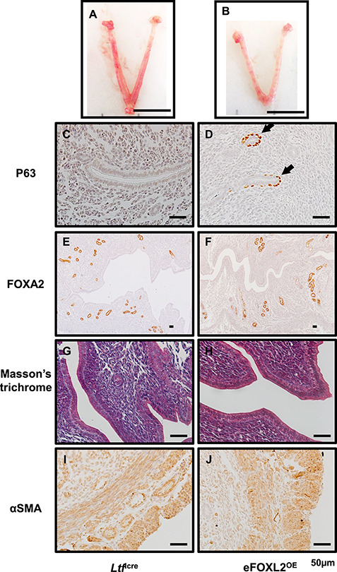
eFOXL2OE exhibited epithelium stratification but no adenogenesis, stroma fibrosis, and myometrial defects. The representative uterine images of Ltficre (A) and eFOXL2OE mice (B) at Pregnancy D3.5. P63 (C and D), FOXA2 (E and F), Masson’s trichrome (G and H), and αSMA (I and J) staining in Ltficre (C, E, G, and I) and eFOXL2OE (D, E, H, and J) uterus. Basal cells with P63 positive staining was detected in the luminal and glandular epithelium of eFOXL2OE uterus. No changes were observed in FOXA2 labeled uterine glands, Masson’s trichrome staining, and αSMA labeled muscle layers. Arrow indicates stratified epithelium. N = 3.
