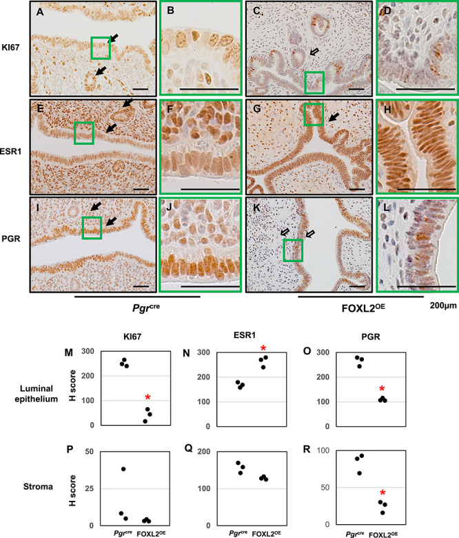
Figure 6.

Protein expressions of KI67, ESR1, and PGR in FOXL2OE uterus at 3 M diestrus stage. Immunohistochemistry of KI67 (A–D), ESR1 (E–H), PGR (I–L) in Pgrcre (A, B, E, F, I, and J), and FOXL2OE (C, D, G, H, K, and L) uterus. H-score of the immunohistochemistry of Ki67 (M and P), ESR1 (N and Q), and PGR (O and R) in the uterine luminal epithelium and stroma showed ESR1 was increased in the epithelium, PGR and KI67 were decreased in the epithelium of the FOXL2OE uterus. Solid arrow indicates cells with strong staining. Open arrow indicates cells with weak staining. N = 3.
