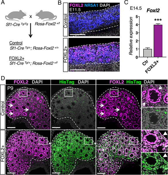
Figure 1.

Constitutive induction of FOXL2 in somatic cells of the ovary. (A) Mouse model for constitutive induction of FOXL2 in the ovarian somatic cells. The Rosa-Foxl2+/f mice were crossed with Sf1-CreTg/Tg mice to produce control and mutant FOXL2+ mice. (B) Immunofluorescence for FOXL2 (magenta), Steroidogenic Factor-1 NR5A1 (blue), and nuclear counterstain DAPI (grey) in control and FOXL2+ ovaries at E11.5. Dotted lines outline the ovaries. Scale bar: 100 μm, n = 4/genotype. (C) qPCR analysis of Foxl2 expression in control and FOXL2+ ovaries at E14.5 The data were analyzed with Student t-test; Bar graphs represent mean ± SEM (n = 6/genotype); ***P < 0.001. (D) Immunofluorescence for total FOXL2 (magenta), ectopic FOXL2 with HisTag (green), and nuclear counterstain DAPI (grey) in control and FOXL2+ ovaries at postnatal day 9 (P9). Dotted lines outline the ovaries. Right panels represent higher magnification images of the outlined area for each single fluorescent channel. Arrows and arrowheads indicate FOXL2+ interstitial cells and FOXL2+ ovarian surface epithelium, respectively. The signal for HisTag in the control ovary came from the autofluorescence of blood cells. Scale bar: 100 μm; n = 4/genotype.
