Figure 4.

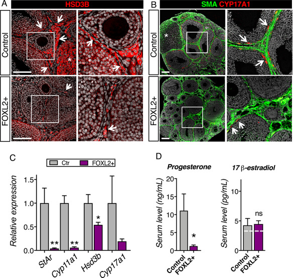
Overexpression of FOXL2 results in defects in steroidogenesis. (A) Immunofluorescence for the steroidogenic enzyme HSD3B in control and FOXL2+ ovaries at 8 weeks of age. Right panels are higher magnification of outlined areas. Arrows point to HSD3B+ theca/interstitial cells. *: HSD3B+ corpus luteum. Scale bar: 100 μm. (B) Immunofluorescence for the steroidogenic enzyme CYP17A1 and alpha smooth muscle actin (SMA) in 8-week-old control and FOXL2+ ovaries. Right panels are higher magnification of outlined areas. Arrows point to CYP17A1+ theca cells in the theca interna. *: corpus luteum. Scale bar: 100 μm; n = 4/genotype. (C) qPCR analysis of steroidogenic enzymes Hsd3b, Cyp17a1, Cyp11a1 and Star expression in control and FOXL2+ ovaries at 8 weeks of age. The results were analyzed with a Mann–Whitney test; mean ± SEM (n = 4–6/genotype); **P < 0.01; *P < 0.05. (D) Serum levels for progesterone and 17β-estradiol in control and FOXL2+ mice at 8 weeks of age. The results were analyzed with Mann–Whitney test; mean ± SEM (n = 6/genotype). The white dotted line represents the detectable range limits for 17β-estradiol immunodetection.
