Figure 5.

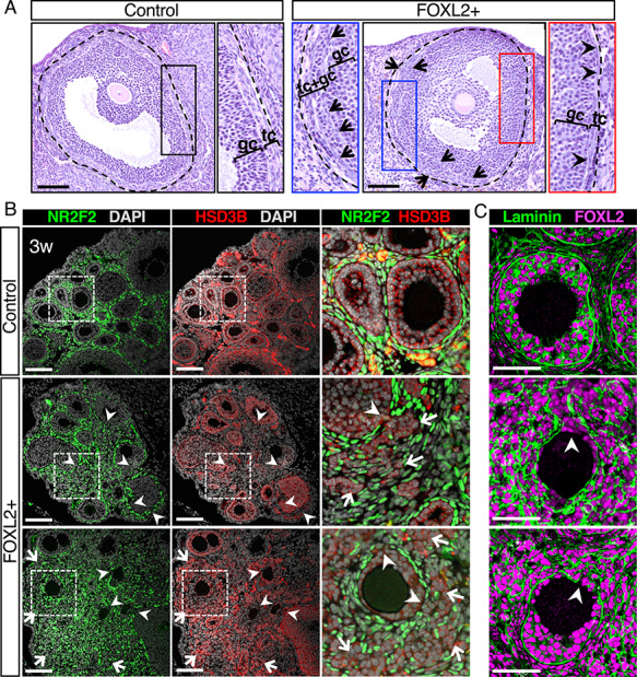
Structural integrity of follicles is compromised in FOXL2+ ovaries. (A) H&E stained antral follicles in adult control and FOXL2+ ovaries. The dotted lines outline the follicles that include the theca interna. Black, blue and red panels represent higher magnification of the respective outlined areas. Arrows indicate flatten theca cells mixed with granulosa cells. Arrowheads point to the theca interna. gc: granulosa cells; tc: theca cells. Scale bar: 100 μm; n = 4/genotype. (B) Immunofluorescence for the interstitial cell marker NR2F2 (green), the steroidogenic enzyme HSD3B, and nuclear counterstain DAPI (grey) in control and FOXL2+ ovaries at 3 weeks of age. The three color-merged panels represent a higher magnification of the outlined square areas. Arrowheads indicate gaps at the junction between granulosa and theca layers. Arrows point to a mixture of granulosa and interstitial cells. Scale bar: 100 μm; n = 4/genotype. (C) Immunofluorescence for FOXL2 and laminin in control and FOXL2+ ovaries at 3 weeks of age. Arrowheads indicate incomplete surrounding of follicles by Laminin in FOXL2+ ovaries. Scale bar: 50 μm; n = 4/genotype.
