
Figure 1.
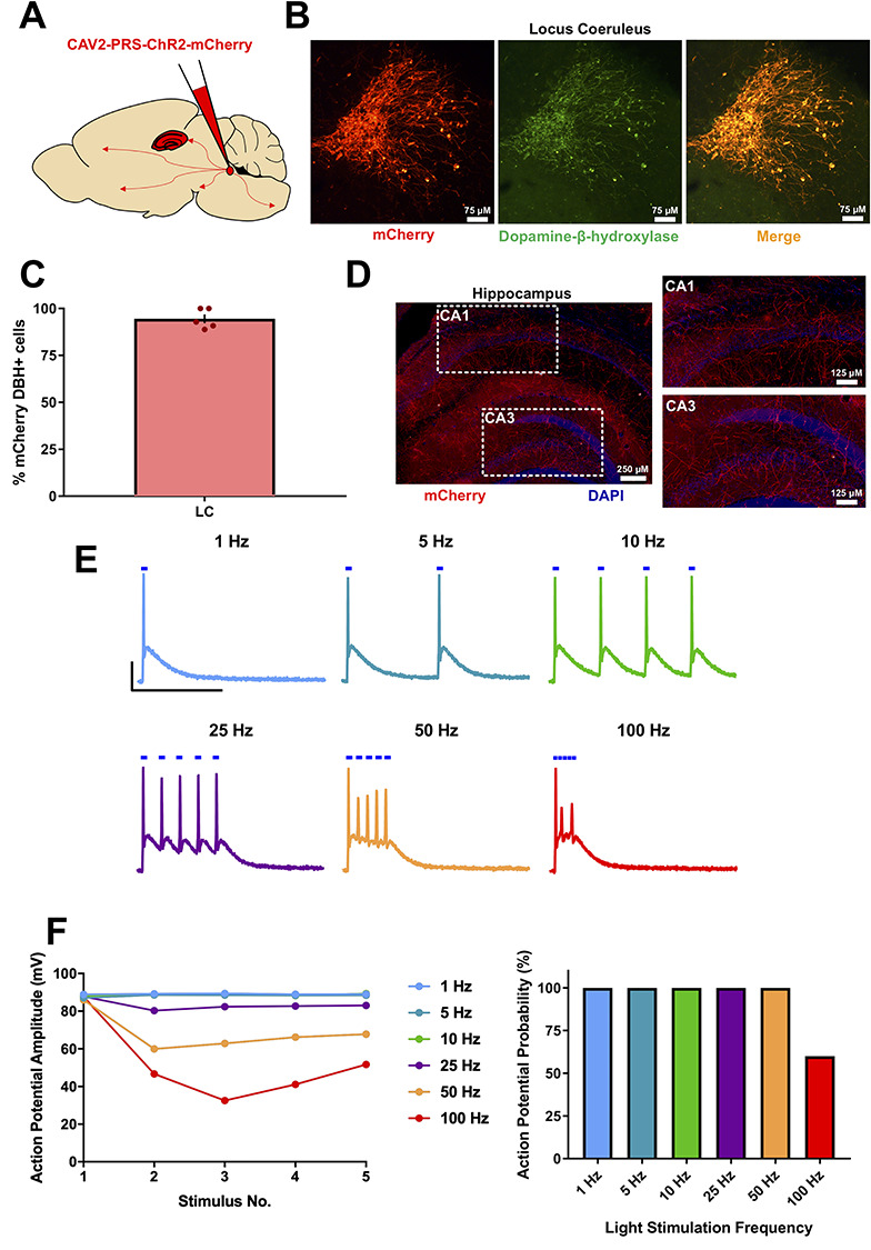
Characterization of the CAV2-PRS-mCherry-ChR2 viral vector expression in mice. (A) Schematic for viral vector injections into the Locus Coeruleus (LC). (B) mCherry fluorescence co-localized with dopamine-β-hydroxylase fluorescence, confirming successful noradrenergic neuronal targeting of the viral vector. (C) % of mCherry+ neurons also showing dopamine-β-hydroxylase expression. (D) mCherry fluorescence in LC fibers within the hippocampus confirming expression of ChR2 in axons. (E and F) Light stimulation of ChR2-mCherry-expressing LC neurons recorded in current-clamp configuration reliably elicited action potentials with stimulation frequencies up to 25 Hz after which amplitude and fidelity began to deteriorate. Scale bars = 25 mV, 200 ms.