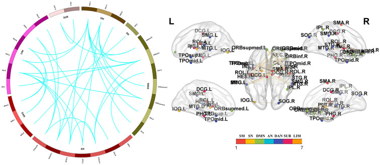
Figure 3.
Functional connectivity network showing decreased functional connections in PD-FOG patients. Compared with the HC group, the PD-FOG group showed a decrease in multiple nodes and connections, which included 29 nodes and 35 connections, mainly involving the sensorimotor cortex (SM), visual network (VN), default mode network (DMN), auditory network (AN) dorsal attention network (DAN), subcortical regions (SUBs) and limbic network (LIM). Network visualization was obtained with BrainNet viewer (http://www.nitrc.org/projects/bnv/) software packages. Abbreviations: PD-nFOG, Parkinson’s disease patients without freezing of gait; HC, healthy control.

