. 2020 Oct 5;10(4):e35. doi: 10.5415/apallergy.2020.10.e35

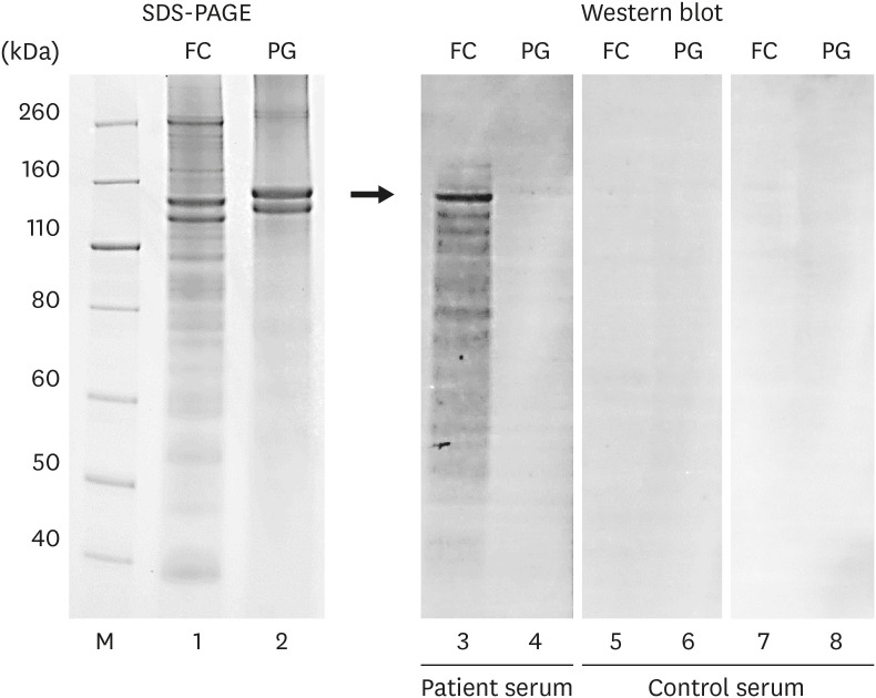
Fig. 1. Interaction between the fish collagen in the gummy tablets and serum from the patient and control. Lanes M, 1, 2, SDS gel stained with Simply Blue; Lanes 3–8, Immunoblot; M, marker; FC, fish collagen in the gummy tablets; PG, pork gelatin; SDS-PAGE, sodium dodecyl sulfate-polyacrylamide gel electrophoresis. Immunoblotting was performed according to the method of Hayashi et al. [10] Proteins were resolved on 4%–12% Bis-Tris SDS gels (Thermo Fisher Scientific, Waltham, MA, USA) and transferred to an Immobilon-P polyvinylidene fluoride membrane (pore size, 0.45 μm; Millipore, Bedford, MA, USA). The membranes were incubated with the patient or volunteer serum and subsequently with a secondary antibody (peroxidase-labeled anti-human IgE [epsilon] antibody; Kirkegaard & Perry Laboratories, Gaithersburg, MD, USA). Bound secondary antibody was detected using the Pierce ECL Plus Western Blotting Substrate (Thermo Fisher Scientific, Waltham, MA, USA). The IgE antibodies in the patient's serum interacted with an approximately 120-kDa protein in the fish collagen sample, but not with pork gelatin.

Fig. 1