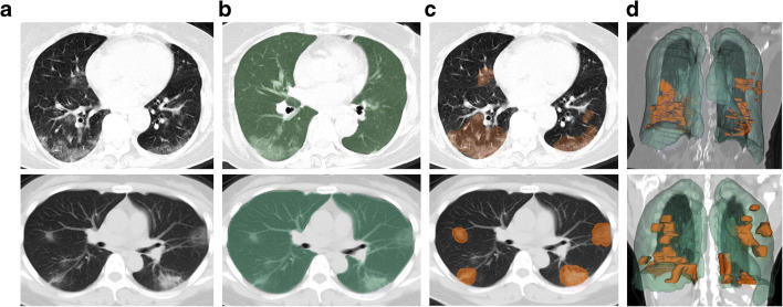
Fig. 3.
Representative chest CT images of two asymptomatic patients. Top—CT scan of a 78-year-old female who never developed COVID-19 symptoms and remained asymptomatic during course of RT-PCR positivity, despite bilateral ground glass opacities on CT. Bottom—CT scan of a 41-year-old female who developed symptoms 5 days after CT. Highlighting the higher attenuation of infiltrates, as consolidations. a Axial chest CT slice with typical infiltrates of COVID-19 pneumonia. b Deep learning–derived whole lung segmentation (green) superimposed over axial chest CT slice. c Superimposed segmented infiltrates (orange) over axial chest CT slice. d Anterior view of 3D volumes of whole lung (green) and infiltrate (orange) segmentations over a coronal chest CT slice

