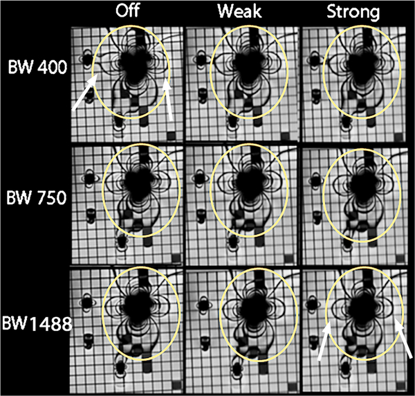
Fig. 3.
In vitro imaging of four metal cardiac devices using steady-state free precession (SSFP) imaging while varying the receiver bandwidth (rows) and asymmetrical echo strength (columns). Ovals represent the size of the artifact in the top left corner, and arrows indicate the changes in artifact size between the first and last images. As bandwidth increases, the image artifacts decrease and image quality decreases. As asymmetrical echo strength increases, the image artifact decreases and image quality decreases. There are significant differences between artifact sizes between the upper left and bottom right panels with similar image quality (arrows). For example, artifact from the bare metal stent and the coil are overlapping in the upper left image and are smaller and separate in the bottom right image

