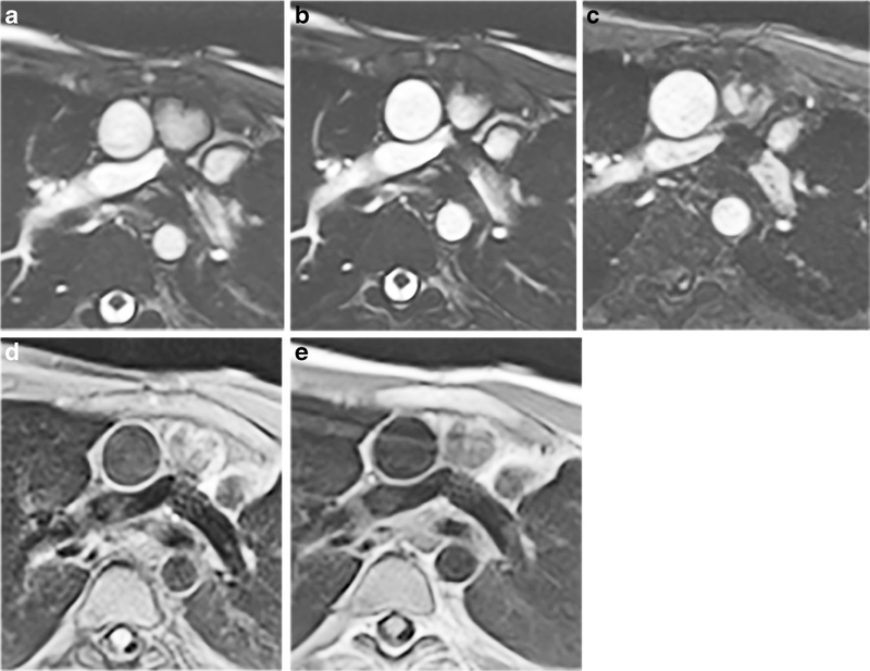
Fig. 8.
Standard vs. sequences optimized for artifact reduction in a 12-year-old boy with a bare metal stent in the proximal left pulmonary artery. Imaging is in the axial plane. a Standard SSFP cine (TR/TE 2.7/1.1 ms, matrix 176×176, FOV 264×264 mm, acceleration factor 2, voxel size 0.8×0.8×6 mm, breath-hold time 12.7 s). b Optimized SSFP cine (TR/TE 2.7/1.1 ms, matrix 176×176, FOV 264×264 mm, acceleration factor 2, voxel size 0.8×0.8×6 mm, breath-hold time 12.6 s. c Optimized GRE cine (TR/TE 3.82/1.69 ms, matrix 176×176, FOV 264×264 mm, voxel size 0.8×0.8×6, acceleration factor 2, breath-hold 16.14 s). d Standard TSE (TR/TE 1,141/31 ms, matrix 192×192, FOV 240×240 mm, voxel size 1.4×1.4×4 mm, breath-hold 12.6 s). e Optimized TSE (TR/TE 1,131/29 ms, matrix 192×192, FOV 240×240 mm, voxel size 1.4× 1.4×4 mm, breath-hold time 12.5 s). In (c), the dephasing artifact decreases significantly with GRE imaging with a receiver bandwidth of 1,488 Hz/pixel, flow compensation off and weak asymmetrical echo. In (d) and (e), improvement in artifact size is seen when when fat saturation is off and bandwidth is increased to 781 Hz/pixel. FOV field of view, GRE gradient recalled echo, SSFP steady-state free precession, TE echo time, TR repetition time, TSE turbo spin echo

