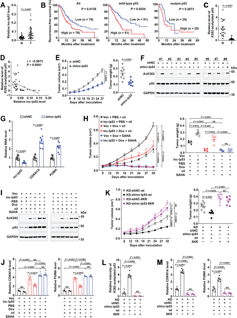
Figure 4.

Lnc‐Ip53 promotes tumor growth and chemoresistance by impeding p53 acetylation in vivo. A) Lnc‐Ip53 was upregulated in human HCC. Lnc‐Ip53 expression was assessed by qPCR in 49 paired HCC (T) and adjacent non‐tumor liver (N) tissues. B) High lnc‐Ip53 level was associated with short recurrence‐free survival (RFS) of HCC with wild‐type but not mutant p53. Data were derived from TCGA. The median lnc‐Ip53 level was chosen as the cut‐off value for separating high‐lnc‐Ip53 group from low‐lnc‐Ip53 group. Survival curves were calculated using Kaplan–Meier method; p‐value was determined by log‐rank test. C,D) The level of K382‐acetylated p53 was C) reduced in HCC and D) inversely correlated with lnc‐Ip53 level. The data of 19 paired T and N tissues were derived from (A) and (C), and applied to Spearman′s correlation coefficient analysis (D). E–G) The xenografts with lnc‐Ip53 silencing displayed E) reduced growth, increase of F) acetylated‐p53 and G) CDKN1A/PUMA. SK‐HEP‐1 cells stably expressing shlnc‐Ip53 or control vector (shNC) were xenografted subcutaneously. n = 8 mice per group. H–J) The xenografts with lnc‐Ip53 overexpression showed H) enhanced growth and chemoresistance, and decrease of I) acetylated‐p53 and J) CDKN1A/PUMA. SK‐HEP‐1 cells stably expressing lnc‐Ip53 or control vector (Vec) were xenografted subcutaneously. PBS and Oil, vehicle controls for Dox and SAHA, respectively. n = 4 mice per group. K–M) Silencing lnc‐Ip53 K) repressed tumor growth and increased L) acetylated‐p53 and M) CDKN1A/PUMA in xenografts with wild‐type p53, but had no effect on those with acetylation‐resistant p53 (8KR). n = 7 mice per group. Acetylated‐p53 was detected by immunoblotting (C,F,I) or immunohistochemistry (IHC; L); CDKN1A and PUMA were examined by qPCR (G,J,M). + or −, cells with (+) or without (−) the indicated treatment. Data are shown as mean ± SEM; p‐values were determined by paired (A,C) or unpaired (E,H,K, right; G,J–M) Student′s t‐test, or two‐way ANOVA (E,H,K, left); NS, not significant.
