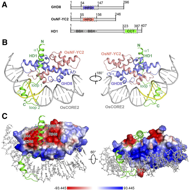
Figure 3.
Overall Structure of the DNA-Bound HD1CCT/GHD8/OsNF-YC2 Complex.
(A) Domain architectures of HD1, GHD8, and OsNF-YC2. The dotted boxes indicate the protein boundaries used for structural determination.
(B) Crystal structure of the DNA-bound HD1CCT/GHD8/OsNF-YC2 heterotrimer. The key secondary structure elements of the HD1CCT are labeled. HD1CCT, GHD8, and OsNF-YC2 are colored green, slate, and salmon, respectively. The DNA OsCORE2 is colored gray with ‘CCACA’ nucleotides highlighted in yellow. Two perpendicular views are presented. All structure figures were prepared with the tool PyMOL.
(C) Electrostatic surface of the GHD8/OsNF-YC2 dimer. Blue, white, and red colors represent positive, neutral, and negative surfaces, respectively. The negative interface of the dimeric GHD8/OsNF-YC2 matches the polar residues in helix α1 of HD1CCT, whereas the positive interface perfectly matches the DNA. The relevant CCT region is shown as cartoon (main chain) and stick (side chains) representations. DNA is shown as gray sticks.

