Figure 4.
Detailed Interactions between the Protein Interaction Element of HD1CCT and GHD8/OsNF-YC2 Dimer.
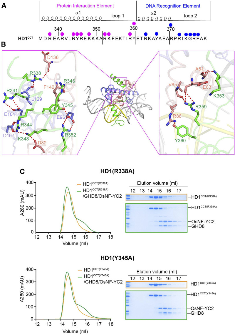
(A) HD1CCT sequence and the key residues interacting with the GHD8/OsNF-YC2 dimer and DNA. HD1CCT consists of two elements: the PIE and the DRE. The magenta dots indicate the residues involved in protein interactions, and the blue dots indicate the residues interacting with DNA.
(B) Zoom-in of HD1CCT residues interacting with GHD8/OsNF-YC2 dimer. HD1CCT, GHD8, and OsNF-YC2 are colored green, slate, and salmon, respectively.
(C) SEC analysis of the interaction between the mutated HD1CCT (R338A or Y345A) and GHD8/OsNF-YC2 dimer. The same eluted fractions of each SEC injection are subjected to SDS-PAGE followed by Coomassie blue staining. CCT domains are fused with an MBP tag at the N terminus. The x axis indicates the elution volume, and the y axis indicates the protein’s absorption at 280 nm. The peak of the MBP-tagged HD1CCT (R338A or Y345A) is colored orange, and the peak of incubation product of MBP-tagged HD1CCT (R338A or Y345A) and GHD8/OsNF-YC2 is colored green. Mutated HD1CCT failed to comigrate forward with GHD8/OsNF-YC2, indicating the abrogation of HD1CCT/GHD8/OsNF-YC2 trimer formation.

