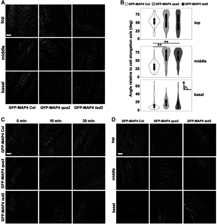
Figure 7.
Cortical Microtubule Patterning Is Altered, and Microtubules Are More Sensitive to Oryzalin Treatment and Mechanical Stress, in qua2 and tsd2 Alleles.
(A) Maximum projections of z-stack images of microtubules from top, middle, and basal hypocotyls of 3-d-old etiolated GFP-MAP4 Col, GFP-MAP4 qua2, and GFP-MAP4 tsd2 seedlings. Bar = 10 μm.
(B) Violin plots of microtubule angles relative to the cell elongation axis in epidermal cells from top, middle, and basal hypocotyls (n ≥ 50 microtubules from at least 12 cells from three seedlings per genotype). The arrow in the small inset indicates the cell elongation axis. **, P < 0.001, Student’s t test.
(C) Cortical microtubule patterning in top hypocotyl regions of 3-d-old etiolated seedlings of GFP-MAP4 Col, GFP-MAP4 qua2, and GFP-MAP4 tsd2 seedlings treated with 10 mM oryzalin over time. Diffuse labeling in the bottom right-hand panels likely represents soluble GFP-MAP4. Bar = 10 μm.
(D) Projections of z-stack images of microtubules from top, middle, and basal hypocotyl regions of 3-d-old etiolated seedlings of GFP-MAP4 Col, GFP-MAP4 qua2, and GFP-MAP4 tsd2 genotypes subjected to a stress of 0.49 N. Bar = 10 μm.

