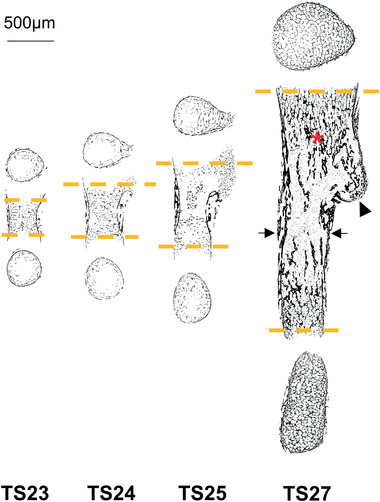
Figure 2.

Evolution of morphology and inner structure of embryonic mice humeri. 2D slices of the µCT reconstructions of one representative sample per age group, showing bone mesoscopic distribution of the mineralized (hard) tissue. Separation into cortical‐ and trabecular‐like structures (arrows and asterisk respectively), as well as mineralization of the deltoid tuberosity (arrowhead) can be observed as early as in TS24. The longitudinal slices were acquired at the center of the rudiment and the cross‐sections at the first slices at each end containing bone structure throughout the whole slice (orange dashed lines).
