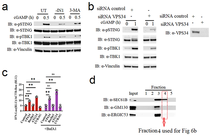
Extended Data Fig. 7. Vps34 Complex I promotes STING activation.
(a) Immunoblot analysis of the indicated proteins from the cell lysates of THP-1 cells pre-treated with DMSO, 10 uM VPS34 inhibitor (VPS34-IN1) or 5 mM 3-MA and then stimulated with 150 nM cGAMP for the indicated time intervals. (n = 3 biologically independent experiments). (b) Immunoblot analysis of the indicated proteins from the cell lysates of HaCaT cells transfected with siRNA-control or siRNA-vps34 for 36 h and then stimulated with cGAMP for the indicated times. (n = 3 biologically independent experiments). (c) Reporter gene assay. HEK-293T with STING stable expression cells were transfected with 50 ng VPS34, Beclin1, ATG14, UVRAG or empty vector, IFNB1 promoter luciferase reporter, and β-actin Renilla reporter. After transfection for 24 h, the cells were treated with/without BafA1 and then stimulated with 200 nM cGAMP for 6 h (n = 3). Statistical analysis of data in panel c was performed using two-tailed one-way ANOVA test (**P = 1.87E-06, ns P = 0.88, **P = 2.46E-09, **P = 0.0063, **P = 0.00053, ns P = 0.10, **P = 1.74E-06, ns P = 0.99, left to right). (d) The isolation of THP-1 cell fractions were obtained by Opti-prep gradient. After collecting the different layers, immunoblot analysis of the indicated proteins was carried out to probe for organelle enrichment of each fraction. The input was from whole cell lysate. Representative blots from two biologically independent experiments with similar results are shown. Data in panel c are shown as means of biological sample triplicates +/- st.dev.

