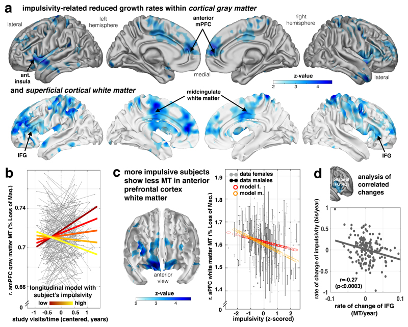
Figure 4. Decreased frontal growth in myelin-sensitive MT in impulsivity.
Myelin marker (MT) in frontal lobe is linked to impulsivity traits. (a) Impulsivity is associated with reduced growth of MT in lateral (inferior and middle frontal gyrus), medial prefrontal areas, motor/premotor and parietal areas in both gray (top panel) and adjacent white matter (bottom panel) depicting negative time/visit by impulsivity interactions (z maps, statistical z-maps, p<.05 FDR and bootstrapping corrected, Supplementary Table 4, one-sided Wald-test, n=497/288 subjects/scans, apply for panels a-c, 51.7% female). (b) Plot shows subjects with higher impulsivity (light yellow) compared to low scoring subjects (dark red) express significantly less MT growth over visits (coloured lines in right panel indicate the interaction effect; y-axis: MT; x-axis: time of scan in years relative to each subject’s mean age over visits). (c) More impulsive subjects show a local decrement in baseline myelin marker (peak middle frontal gyrus, p<0.05, FDR and bootstrapping corrected, Supplementary Table 5) in lateral and orbitofrontal areas (fixed for other covariates, e.g. time/visits, mean age of subject, sex). Right panel shows the plot of MT in this peak voxel over impulsivity (x-axis, z-scored) and with adjusted data (gray/black) and model predictions (red/orange, effects of interest: intercept, impulsivity, sex by impulsivity). (d) Bilateral IFG not only shows a reduced myelination process for higher impulsivity (as shown in a, b), but this reduced growth rate is more strongly expressed in subjects who manifest an accentuated impulsivity growth over study visits, such that subjects who manifest an even more restricted growth in myelin become more impulsive (Pearson’s correlation and p-value from corresponding t-distribution based on n=188 independent subjects with available follow-up scans).

