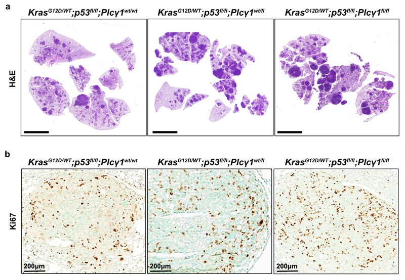
Extended Data Fig. 9. PLCγ1 deletion accelerates lung tumorigenesis in mice.
(a) Representative hematoxylin & eosin (H&E) staining of LSL-KrasG12D/WT;p53flox/flox;Plcγ1wt/wt, LSL-KrasG12D/WT;p53flox/flox;Plcγ1wt/flox and LSL-KrasG12D/WT;p53flox/flox;Plcγ1flox/flox mouse lung sections, 10 weeks after Cre induction. Scale bar: 5000μm.
(b) Representative immunohistochemistry images showing Ki67-positive cell staining in mouse lung tumors from LSL-KrasG12D/WT;p53flox/flox;Plcγ1wt/wt, KrasG12D/WT;p53flox/flox;Plcγ1wt/flox and LSL-KrasG12D/WT;p53flox/flox;Plcγ1flox/flox mice 10 weeks after Cre induction. This is related to main Fig. 7e.

