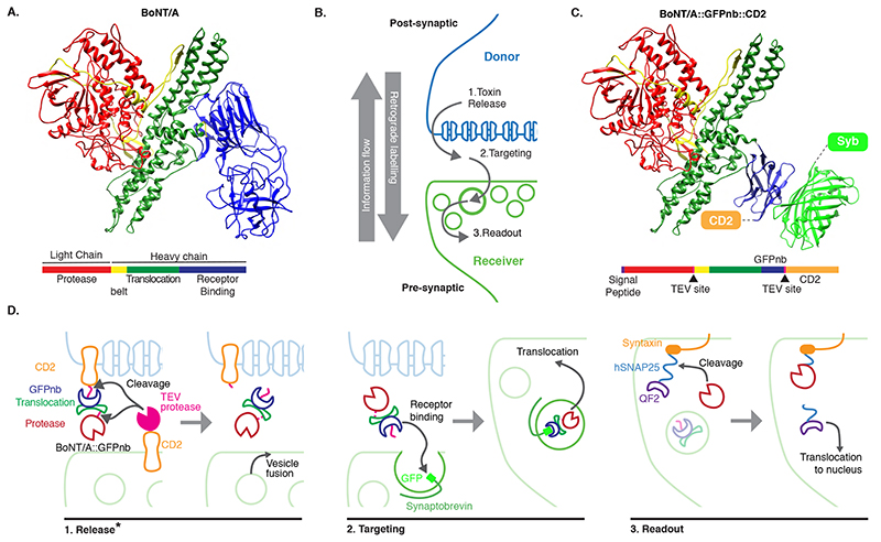
Figure 1. System design.
(A) Molecular structure of BoNT/A (PDB id 3BTA, [40]). Toxin domains are colored red (protease), yellow (belt), green (Translocation, TD) and blue (Receptor Binding, RBD). (B) Schematic showing the steps leading to labelling. (C) Retargeted BoNT/A: the RBD from (A) is replaced by an anti-GFP nanobody (blue). Also shown in light green the bound GFP (PDB id 3OGO, [13]). (D) Proteins and molecular mechanisms mediating each step from (B). Colors as in (C) except pink sections in BoNT/A::GFPnb::CD2 which indicate TEV cleavage sites. * The release step was shown to be TEV independent later in the project.

