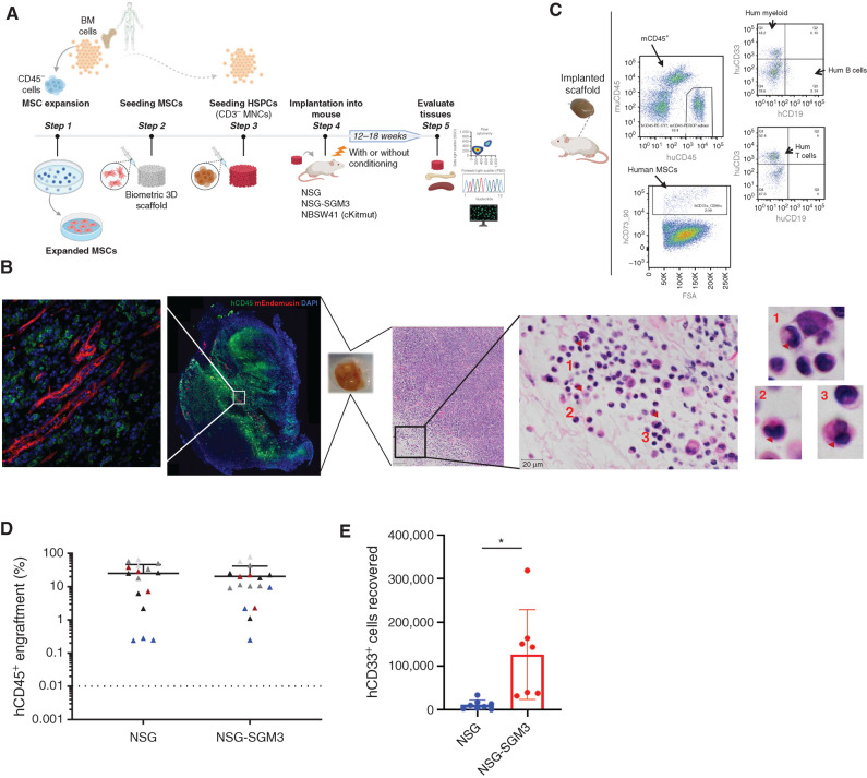
Figure 1.
Humanized scaffold system in NSG and NSG-SGM3 immunodeficient mice. A, Schematic representation of the in vivo protocol used for generating humanized scaffolds. Illustration was created with BioRender.com. B, Representative example of immunofluorescence and hematoxylin and eosin (H&E) staining for the scaffold retrieved following the xenotransplantation. H&E staining of a section of the engrafted scaffold shows multiple neutrophils with hypolobated nuclei (examples indicated with arrowheads), consistent with dysplasia in the granulocytic lineage. C, Representative flow cytometry plot of the cells retrieved from the humanized scaffolds following xenotransplantation. D, Comparison of hCD45+ cells engraftment in humanized scaffolds retrieved from NSG and NSG-SGM3 mice 12 weeks following the scaffold implantation. E, Absolute cell counts of human myeloid (hCD45+hCD33+) cells retrieved from humanized scaffolds implanted in NSG and NSG-SGM3 mice. *, P ≤ 0.05.

