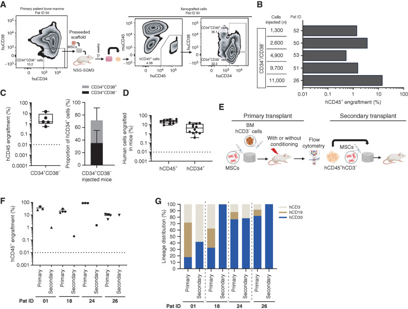
Figure 3.
MDS-SCs engraft in humanized niches in NSG-SGM3 mice and demonstrate long-term self-renewal capacity. A, Representative flow cytometry plot showing the day 0 patient BM CD34/CD38 phenotype and engraftment of hCD45+ cells in humanized scaffolds as well as the hCD34/hCD38 phenotype of these xenografted cells in NSG-SGM3 mice. Pat ID, patient ID. B, Total hCD45+ cell engraftment in humanized scaffolds that were seeded with CD34+CD38− MDS-SCs in NSG-SGM3 mice. C, Percentage of hCD45+ cells (left) in mice injected with CD34+CD38− MDS cells and proportion of CD34+C38−/CD34+CD38+ cells within hCD34+ HSPCs in these humanized scaffolds retrieved from NSG-SGM3 mice. D, Percentage of hCD45+ cells and proportion of hCD34+ cells within the hCD45+ cell fraction in the humanized scaffolds that were seeded with MDS CD3− MNCs. E, Schematic representation of the in vivo protocol for secondary transplantation. F, Percentage of total hCD45+ cells engrafted in humanized niches from primary and secondary NSG-SGM3 mice. G, Lineage distribution within the hCD45+ cells in humanized niches implanted in primary and secondary NSG-SGM3 mice. Mice were kept alive for up to 18 weeks following the scaffold implantation.

