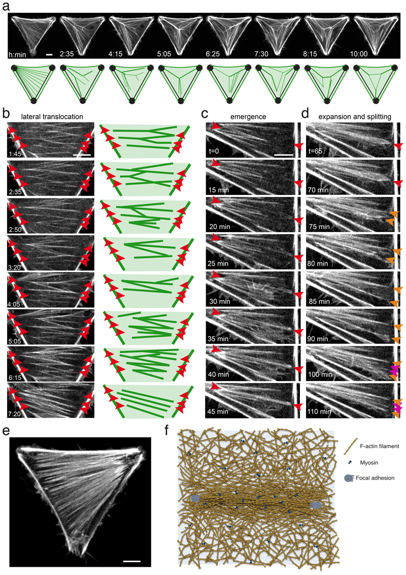
Figure 6. Emergence and translocation of cytoplasmic bundles in the cortical meshwork.
a. Live imaging of RPE1-LifeAct-GFP cells on tripod-shaped micropattern showing the global and permanent remodelling of network architecture, suggestive of a complex interplay of longitudinal and lateral forces on cytoplasmic bundles. See corresponding Video 5. N=1 experiment. Scale bar = 10 µm.
b. Live imaging of RPE1-LifeAct-GFP cells on tripod-shaped micropattern highlighting network reconfiguration by lateral translocation of cytoplasmic bundles in the absence of anchorage displacement (red arrows). See corresponding Video 6. N=1 experiment. Scale bar = 10 µm.
c. Live imaging of RPE1-LifeAct-GFP cells on tripod-shaped micropattern revealing the emergence of cytoplasmic bundles from the cortical meshwork (in between red arrows). See corresponding Video 7. N=1 experiment. Scale bar = 10 µm.
d. Live imaging of RPE1-LifeAct-GFP cells on tripod-shaped micropattern showing the lateral expansion of a bundle (red arrow), its splaying into a wider structure (orange arrows) and its re-coalescence into several adjacent bundles (magenta arrows). See corresponding Video 7. N=1 experiment. Scale bar = 10 µm.
e. RPE1-LifeAct-GFP cells on tripod-shaped micropattern displaying a dense and quasi-continuous network of cytoplasmic bundles. N=1 experiment.
In all panels, time is indicated in hours and minutes. Scale bar = 10 µm.
f. Schematic representation of the stress fiber anchored at its two edges on the substrate via focal adhesions (blue disks) as a fully embedded structure within the surrounding contractile actin cortex (myosins are represented by blue bow-ties).

