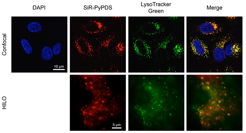
Extended Data Fig. 8. SiR-PyPDS mainly accumulates in lysosomes.
Representative confocal and HILO microscopy images obtained in the presence of SiR-PyPDS (1 μM in confocal and 40 nM in HILO) and LysoTracker Green (50 nM), confirming co-localisation of extranuclear staining with lysosomes. Experiments have been repeated 3 times providing similar results.

