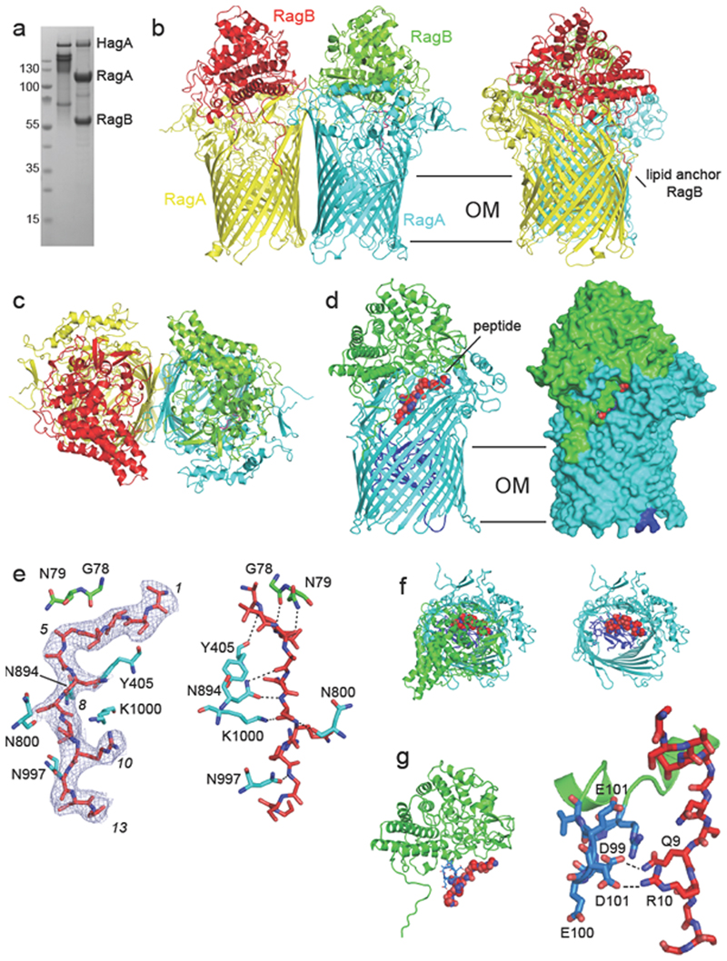
Figure 1. Crystal structure of the RagA2 B 2 transporter suggests bound peptides.
a, SDS-PAGE gel showing purified RagAB from W83 KRAB before (left lane) and after boiling in SDS-PAGE sample buffer. The gel is representative of three independent purifications. b,c, Views of RagA2B 2from the plane of the OM (b) and from the extracellular space (c). d, Side views of RagAB (right panel; surface representation) showing the bound peptide as a red space filling model. The plug domain of the RagA TBDT (cyan) is dark blue. e, 2Fo-Fc density (blue mesh; 1.0 σ, carve = 1.8) of the peptide after final refinement (left panel). The right panel shows potential hydrogen bonds (distance < 3.6 Å) between RagAB and the peptide backbone. Arbitrary peptide residue numbering is indicated in italics. f, Extracellular views of RagAB with (left panel) and without the RagB lid (green). g, Side view (top panel) and close-up of the RagB acidic loop (blue) and the bound peptide. Potential hydrogen bonds are indicated by dashed lines. Structural figures were made with Pymol69.

