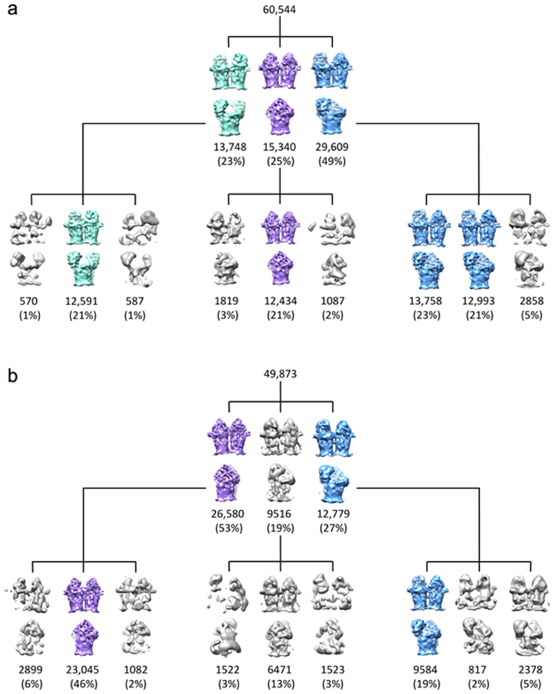
Extended Data Fig. 10. Cryo-electron microscopy data support substrate-induced lid closure in RagAB.
a,b, 3D classes for RagAB ‘as purified’ (a) and in the presence of 50-fold excess P21 peptide (b). Classes corresponding to the CC, OC and OO states are coloured purple, blue and green respectively. Junk or ambiguous classes e.g. where RagA barrels are incomplete are coloured grey. In the presence of P21 there was no clear OO state whilst the the proportion of the CC state increased, supporting the proposed mechanism of substrate capture.

