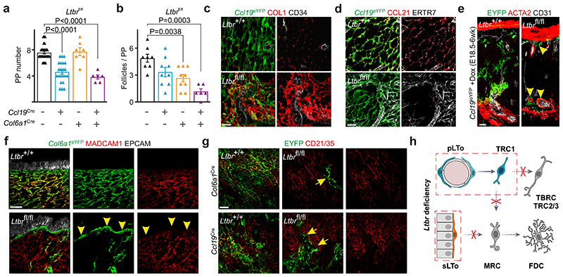
Figure 3. Lymphotoxin-β-receptor signaling dependent FRC organization in PP microdomains.
a-b, Number of PPs and B cell follicles in PPs from adult Ltbr fl/fl (n=30), Ccl19 Cre Ltbr fl/fl (n=22), Col6a1 Cre Ltbr fl/fl (n=7) and Ccl19 Cre Col6a1 Cre Ltbr fl/fl (n=6) mice. c-d, Confocal microscopy analysis showing EYFP+ FRC networks in adult PP. e, Images depict EYFP+ cells representing progeny of E18.5 progenitors in the T cell zone of adult PP of inducible Ccl19 ieYFP mice. Arrowheads indicate perivascular location of Ltbr-deficient EYFP+ cells. f, Confocal microscopy of EYFP+ FRC networks in subepithelial dome and g, B cell follicle, arrowheads in f indicate subepithelial YFP+ cells, arrows in g indicate EYFP+CD21/35- cells. h, Schematic depiction of LTβR signaling in FRC differentiation. Scale bars, 20 μm (c-e,g), 30 μm (f),. Images are representative of 3-4 mice per group, at least 2 independent experiments. (a,b) SEM is indicated; P values as per two-tailed Mann Whitney Test.

