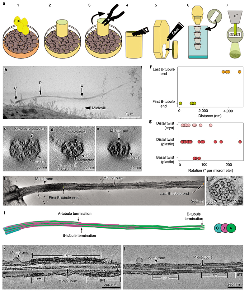
Fig. 1. I Room temperature ET of MDCK-II primary cilia.
a, Description of the steps for resin-embedding and serial sectioning of MDCK-II monolayers. b, Montage of projections from five serial sections covering the entire primary cilium. C, D and E are the areas of the cilium that were digitally sectioned to obtain the images shown in panels c,d and e, respectively. c-e, Representative proximodistal tomographic slices from the same cilium shown in b, showing the axonemal organization of microtubule doublets and singlets. c, Cross-section of the cilium proximal segment showing that 9-fold symmetrical arrangement of the microtubule doublets is already lost. d,e, The number of microtubule doublets (d) and microtubules singlets (e) decreases in the distal part of the cilium. f, Distance measured from the distal side of the basal body to the first and last B-tubule terminations. g, Axonemal twist measured at the basal and distal regions of the cilium. H,i, Tomographic section along a cilium (H), and its microtubule segmentation (i), showing representative positions of first and last B-tubule terminations. J, Ciliary cross-section showing the variety of axonemal structures other than microtubules. k,l, Longitudinal sections through cilia containing IFT train-like particles sandwiched between the ciliary membrane and microtubule singlets. Asterisks indicate locations of unidentified densities.

