Figure 2.
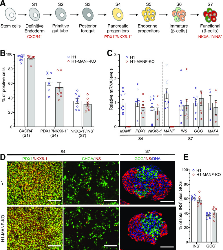
MANF KO hESC differentiate normally to β-cells. A: Schematic presentation of the seven-stage differentiation protocol (for further details, see research design and methods). B: Graphical presentation of flow cytometry analysis of the differentiating hESC for specific relevant markers of each stage in H1 (WT) and H1-MANF-KO (N = 8 for S1 and S7 cells; N = 7 for S4 cells). For the definitive endoderm stage, CXCR4 was used as a surface marker, PDX1 and NKX6-1 double-positive staining was used as a marker of the pancreatic progenitor population (S4), while NKX6-1 and insulin (INS) double-positive staining was used as a marker of end-stage β-like cells (S7). C: Relative gene expression levels of pancreatic genes analyzed by RT-qPCR in H1 and H1-MANF-KO (N = 4–9). D: Immunocytochemistry analysis of S4 monolayer cells for PDX1, NKX6-1, chromogranin A (CHGA), and insulin (INS) (scale bars = 100 μm) and S7 aggregates for INS, glucagon (GCG), and the nuclear stain Hoechst (N = 6) (scale bars = 100 μm). E: Quantification of the percentage of INS-positive and GCG-positive cells in H1 and H1-MANF-KO SC-islets. H1 is presented as blue circles, and H1-MANF-KO is presented as red circles. Data are presented as mean ± SEM.

