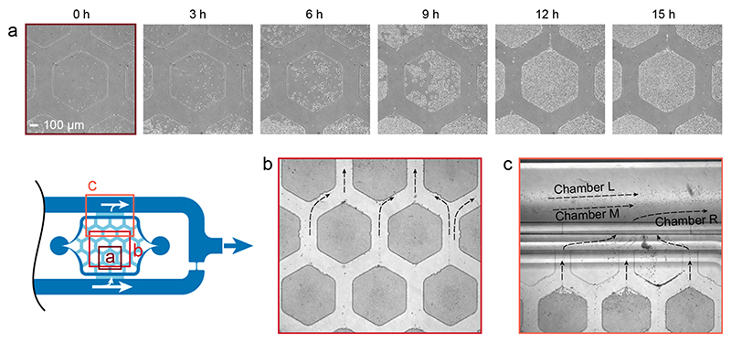
Figure 6. Cell growth and wash away.
(a) Phase contrast time-lapse micrographs (Plan Fluor 10X Ph1) of S. cerevisiae cells, loaded at a density of 5 × 10⁷ cells/mL, and their proliferation until complete pad filling after 15 h (see Video S-1 for full time-lapse movie). (b) Overgrowing cells are washed away by the constant medium flow between the hexagonal pads (bright field micrograph, Plan Fluor 4X). Laminar streamlines minimize cell crosstalk between different pads (see also real-time Video S-2). (c) Cells leaving the cell culturing area exit the chip through the perfusion channel surrounding the culturing chambers (bright field micrograph, Plan Fluor 4X). The parallel chamber arrangement prevents cells from one cell

