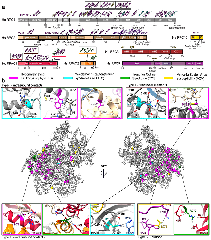
Fig. 5. Disease-associated mutations of Pol III.
a, Schematic representation of Pol III subunits affected by disease-associated mutations mapped onto the domains. R279Q/W mutations (starred) associated with TCS17 was also reported in individuals with HLD10. A – anchor; N – N-terminal domain; C – C-terminal domain; DM – dimerization domain; WH – winged-helix; CC – coiled-coil. b, Disease-associated mutations (coloured spheres associated with each disease) mapped on a cartoon representation of human Pol III. Close up views of representative mutations belonging to one of 4 identified types (see Methods): I – intrasubunit contacts, II – functional elements, III – intersubunit contacts and IV – surface are shown. Grey solid lines - contacts between residues, black dotted lines - hydrogen bonds.

