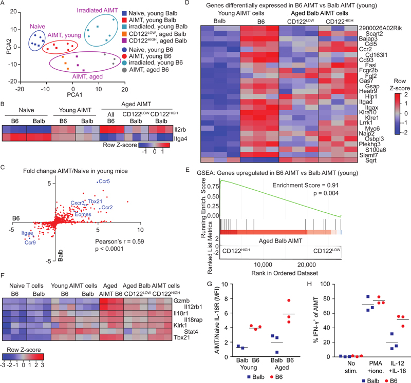
Figure 3. Similar gene expression profiles of thymic and peripheral AIMT cells.
(A-E) Naïve (CD44-) and/or AIMT (CD44+ CD49d-) CD8+ T cells were sorted from lymph nodes and spleens from young (5-12 weeks) untreated or young irradiated (4 Gy, 3 weeks) B6 and Balb or aged B6 mice (76 weeks). CD122HIGH and CD122LOW AIMT cells were sorted from lymph nodes and spleens from aged Balb mice (54-55 weeks). The transcriptome of these populations was analyzed by RNA sequencing.
(A) PCA analysis of the gene expression profiles of the individual samples normalized to avoid the strain-specific differences (top 500 variable genes).
(B) Relative expression (z-score) of Il2rb (CD122) and Itga4 (CD49d) in individual samples using normalization for strain-specific differences.
(C) Ratios of transcript levels in AIMT cells to naïve cells (young mice) were calculated for each gene and plotted as log2 for B6 and Balb strains. Selected genes are indicated. The correlation was calculated using Pearson’s correlation coefficient.
(D) Relative expression (z-score) of genes showing significantly different expression between AIMT cells from young B6 and Balb mice (after the normalization for strain-specific differences) in AIMT cells from B6 and Balb mice and in CD122HIGH and CD122LOW AIMT cells from aged Balb mice.
(E) The gene set enrichment analysis (GSEA) of genes with significantly higher expression in B6 than in Balb mice (shown in Fig. 3D) in CD122HIGH vs. CD122LOW AIMT cells from aged Balb mice.
(F) Relative expression (z-score) of selected genes involved in by-stander cytotoxicity between AIMT cells from young B6 and Balb mice (after the normalization for strain-specific differences) in AIMT cells from B6 and Balb mice and in CD122HIGH and CD122LOW AIMT cells from aged Balb mice.
(G) Expression of IL-18R on the surface of AIMT cells from the lymph nodes of young and aged Balb and B6 mice measured by flow cytometry. Geometric mean fluorescent intensity (MFI) was normalized to naïve cells in the same sample. Mean is shown. n = 3 independent experiments.
(H) Production of IFN-γ by AIMT cells (gated as CD8α+ CD44+ CD49d+) from young adult B6 or Balb mice was measured by flow cytometry. Lymph node cells were stimulated with PMA + ionomycin or with IL-12 and IL-18 or left unstimulated for 8 hours and analyzed by flow cytometry. n = 3 independent experiments.

