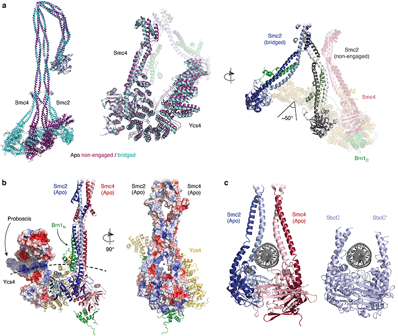
Extended Data Fig. 5. Conformation al changes and putative DNA binding sites in the apo condensin complex.
a, Structural alignment of pseudo-atomic models of overall apo complexes in the non-engaged and bridged states. b, Electrostatic potential maps of Ycs4 (left) and the Smc2–Smc4 heads and coiled coils (right); red: –5 keT, blue: +5 keT. The dotted line indicates a putative path for a DNA double helix that goes through the coiled coils above the ATPase heads and also includes the positively charged patch on Ycs4 near the ‘proboscis’ protrusion. c, Placement of a DNA double helix in the space between the neck regions of the Smc2 and Smc4 coiled coils in the apo non-engaged state, based on a comparison to a DNA co-crystal structure of the E. coli SbcC (Rad50) head dimer in the engaged state (PDB code 6S85).

