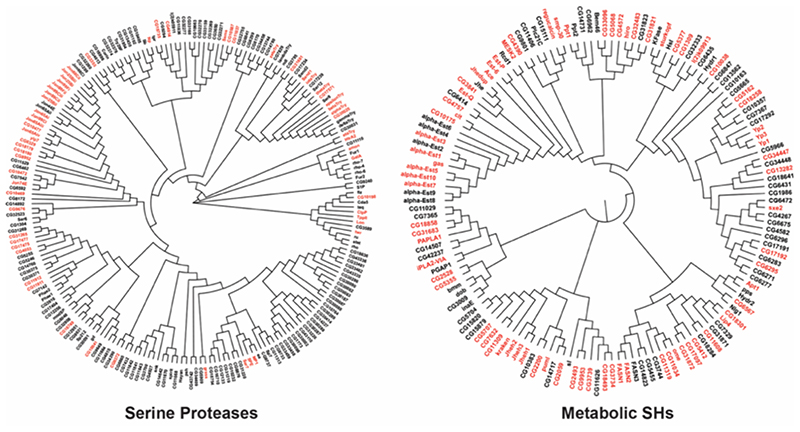
Figure 7. A chemoproteomic atlas of SH activities in Drosophila melanogaster.
A dendrogram analysis of all the predicted serine proteases and metabolic SHs in Drosophila melanogaster, in which, the red and black coloring represents enzymes that were enriched and not enriched respectively in the LC-MS/MS based ABPP experiments using the SH-directed FP-biotin probe. All protein sequences of the respective category (serine proteases and metabolic SHs) were aligned using the MUSCLE program of the MEGA-X software by the neighbor joining method, where the branch length denotes relatedness between protein sequences.

