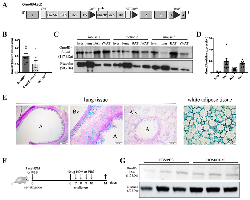
Figure 1. Ormdl3-LacZ reportermice as a useful tool to study ORMDL3 expression.
A)Ormdl3-LacZ reportermice (Ormdl3 LacZ/LacZ) were generated by inserting a targeting construct of the EUCOMM consortium into the first intron of Ormdl3. Exon 2, exon 3 and part of exon 4 are flanked by two loxP sites, which enable the generation of conditional knockout mice upon crossing with mice expressing Cre-recombinase.
B)Ormdl3 mRNA expression levels in lungs from Ormdl3+/+, Ormdl3 LacZ/LacZ and Ormdl3-/- mice. Expression values are shown relative to means of the wildtype group. Data were pooled from 2 experiments (n=7,6,4; means +/-SEM).
C)Western blot showing β-galactosidase expression in liver, lung, brown adipose tissue (BAT) and white adipose tissue (WAT) in three individual Ormdl3-LacZ reportermice. β-tubulin was used as a loading control.
D)Ormdl3 transcript levels in lung, BAT, WAT and liver in wildtype mice. Expression values are shown relative to means of lung samples (means +/- SEM).
E)Immunohistochemistry analysis of β-galactosidase expression (blue) on lung OCT-inflated cryosections and WAT of Ormdl3-LacZ reportermice. Periodic-acid Schiff staining was used as counterstaining. A = airway; Bv = blood vessel; Alv = alveoli.
F)Scheme representing the acute house dust mite (HDM)-dependent asthma model.
G)Western blot showing β-galactosidase expression in lung tissue from mock- and HDM-challenged Ormdl3-LacZ reportermice.

