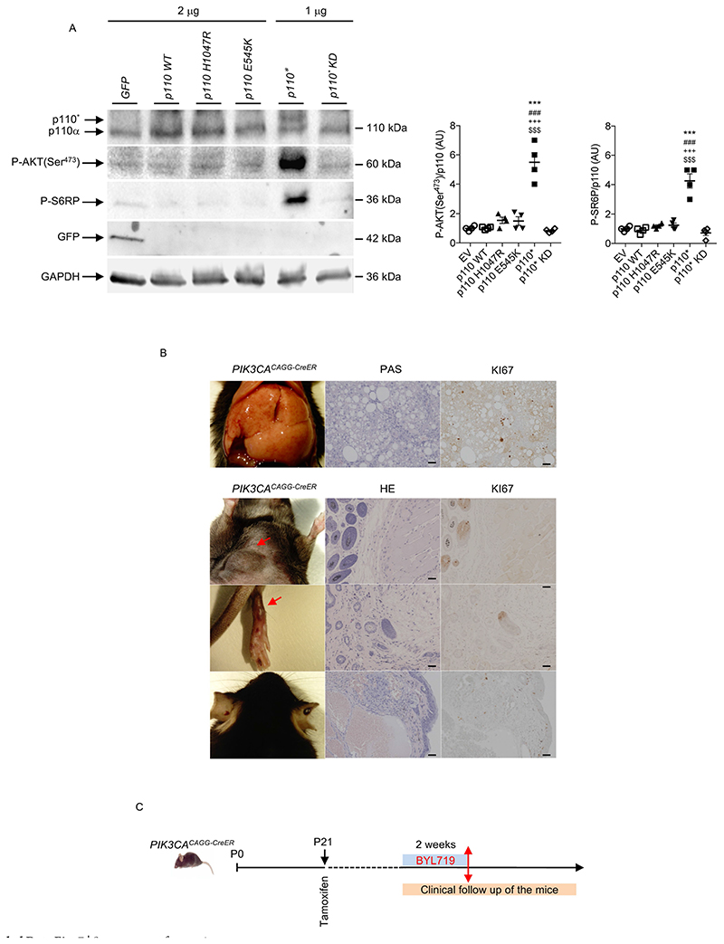
Extended Data Fig. 7. Recruitment of the AKT/mTORC pathway by the different forms of mutant p110.
a, Western blot and quantification of p110, P-AKT (Ser473), P-S6RP and GFP in HeLa cells transfected with plasmids containing cDNA encoding p110*, p110* kinase-dead mutant (p110* KD) as a control, H1047R mutation or E545K mutation. Cells transfected with the p110* mutant showed a more powerful effect on the activation of the AKT/mTORC pathway than the others (n = 4 independent experiments). All data are shown as mean ± s.e.m. ANOVA followed by Tukey–Kramer test (two-tailed). p110* versus H1047R mutation, ***P < 0.001. p110* versus E545K mutation, ***P < 0.001. p110* versus wild-type p110, +++ P < 0.001. p110* versus p110* KD, $$$P < 0.001. Negative control is a vector that contains cDNA encoding GFP. b, Histological examination of different tissues from PIK3CACAGG-CreER mice. Left column, from top to bottom, liver, abdominal tumour, leg and ear abnormalities. Middle column, PAS or HE staining of the tissue. Right column, Ki67 staining of the same tissue (n = 8 mice). Scale bars, 20 μm. c, Design of the experiment shown in Fig. 2h. PIK3CACAGG-CreER mice received a single dose of 4 mg kg−1 tamoxifen and were followed for one month. Once the tumours became visible, BYL719 was started for two weeks and then withdrawn.

