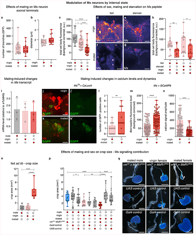
Extended Data Fig. 6. Post-mating modulation of Ms neurons.
a-b, Analysis of Ms neuron crop terminals in virgin and mated females. Neither the number of axonal branches (a) nor their diameter (b) is significantly different between virgin and mated females. c, Quantifications of Ms staining levels in the cell bodies of PI neurons of wild-type, ad libitum-fed males, virgin females and mated females. Mated females have less Ms peptide than virgin females or males; virgin females have less peptide than males. d-h, Comparison of Ms peptide levels in the cell bodies of PI neurons in fed versus starved virgin and mated females. Representative images of Ms staining in the cell bodies of the PI neurons of fed virgin females (d), starved virgin females (e), fed mated females (f) and starved mated females (g). h, Quantification of Ms staining in the cell bodies of PI neurons shows that Ms levels are reduced in mated females compared to virgins, irrespective of fed or starved status. i, RT-qPCR expression data for Ms transcript levels in the brain of ad libitum-fed, control males (grey column), virgin females (pink column) and mated females (red column). No significant differences are apparent between groups. j-l, CaLexA-based assessment of mating-triggered changes in PI Ms neuronal activity, achieved by adult- and Ms-confined CaLexA expression (MsTS > CaLexA). Representative images of ad libitum-fed, wild-type virgin (j,j’), and mated females (k,k’) are shown. Ms neurons are labelled with anti-Ms antibody (in red) and CaLexA channel is shown as a single channel (in green), for clarity. l, Quantification of CaLexA-derived GFP-positive cells in PI Ms neurons of virgin (pink box) and mated (red box) females, showed that fewer cells are CaLexA-positive in virgin compared to mated females; each data point corresponds to a different brain. m,n, Quantification of baseline GCaMP fluorescence (corrected for background) (m) and amplitude of GCaMP fluorescence oscillations (n) in the cell bodies of PI Ms neurons of virgin females (pink box) or mated females (red box). Each data point corresponds to an individual cell measurement. Higher GCaMP signal and reduced oscillation amplitude are detected in mated females. o, Crop area quantifications in wild-type, ad libitum-fed males, virgin females and mated females. The crop of mated females is bigger than that of virgin females or males. p,q, Effects of sex and mating status on Ms signalling contribution to crop size. p, Quantification of crop area upon adult-specific downregulation of MsR1 in visceral muscles shows that this was significantly reduced in mated females but not in males or virgin females, as compared to respective controls. q, Representative crop images of genotypes quantified in m. Scale bars: d-g and j-k’= 20μm and q = 500μm. See Supplementary Information for a list of full genotypes, sample sizes and conditions. In all boxplots, line: median; box: 75th-25th percentiles; whiskers: minimum and maximum. All data points are shown. *: 0.05>p>0.01; **: 0.01>p>0.001; ***: p<0.001.

