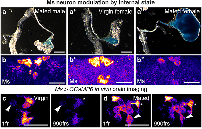
Fig. 2. Reproductive modulation of Ms neurons.
a-b″, Representative dissected intestines (top) and Ms stainings of the PI region of the brain (bottom) of wild-type flies. Mated females have more expanded crops (a″) and less Ms in their cell bodies (b″) than virgin females (a’,b’) or mated males (a,b). In b-b″, fluorescence signals are pseudo-coloured; high to low intensity is displayed as warm (yellow) to cold (blue) colours here and thereafter. c,d, Temporally defined video snapshots of Ms-driven GCaMP6 activity in the PI of virgin (c), or mated (d) females, imaged over 1000 frames (frs, each frame acquired every 427 milliseconds). Asterisks and arrows highlight two randomly chosen Ms neurons. Scale bars = 20μm except for a-a″ = 500μm.

