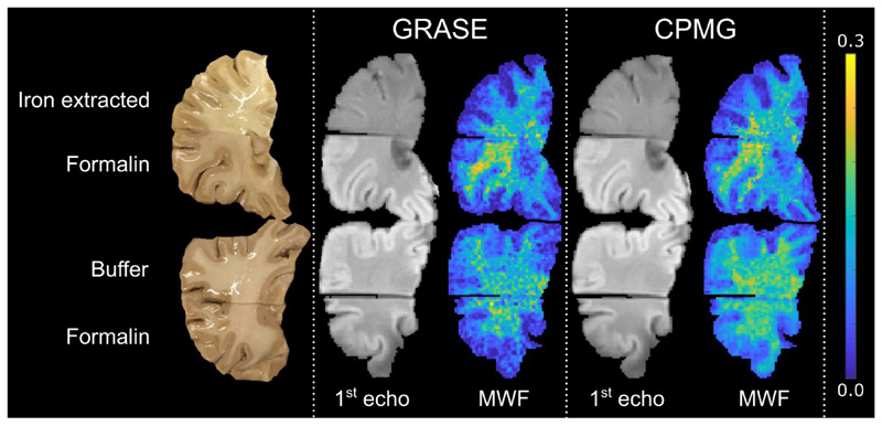
Fig. 3.
The top row shows a brain slice, the corresponding images of the 1st echo (TE = 10 ms) and MWF maps assessed using the GRASE and CPMG sequence, respectively. The brain slice was cut into two parts, where the upper part underwent 12 days of iron extraction and the lower part was kept in formalin as reference. A clear change in image contrast and decrease in MWF is observed in the iron extracted part compared to the reference part. The bottom row shows a second brain slice, out of the same brain, the corresponding images of the 1st echo (TE = 10 ms) and MWF maps for both the GRASE and CPMG sequence. The brain slice was cut into two parts where the upper part was kept in the buffer solution without iron chelator and the lower part in formalin for 12 days. No change in image contrast and MWF was observed in the buffer treated part compared to the part stored in formalin.

