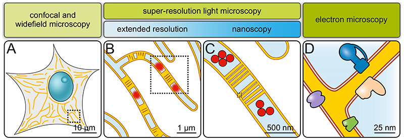
Fig. 3. Imaging mitochondria across scales.
A) Diffraction-limited microscopy (confocal or widefield microscopy). Overall mitochondrial morphology and network dynamics can be visualized. Only limited information on sub-mitochondrial protein distributions. B) Extended-resolution microscopy (diffraction-limited super-resolution microscopy). Network dynamics, but also inner mitochondrial dynamics. Groups of cristae and, under some conditions, single cristae are visible. Mitochondrial sub-compartments can be analyzed. C) Nanoscopy (diffraction-unlimited super-resolution microscopy). Detailed sub-mitochondrial protein distributions and individual cristae can be resolved. D) Electron microscopy. Precise membrane architecture and lipid bilayers are resolved. With specific approaches, protein distributions and even protein structures can be determined.

