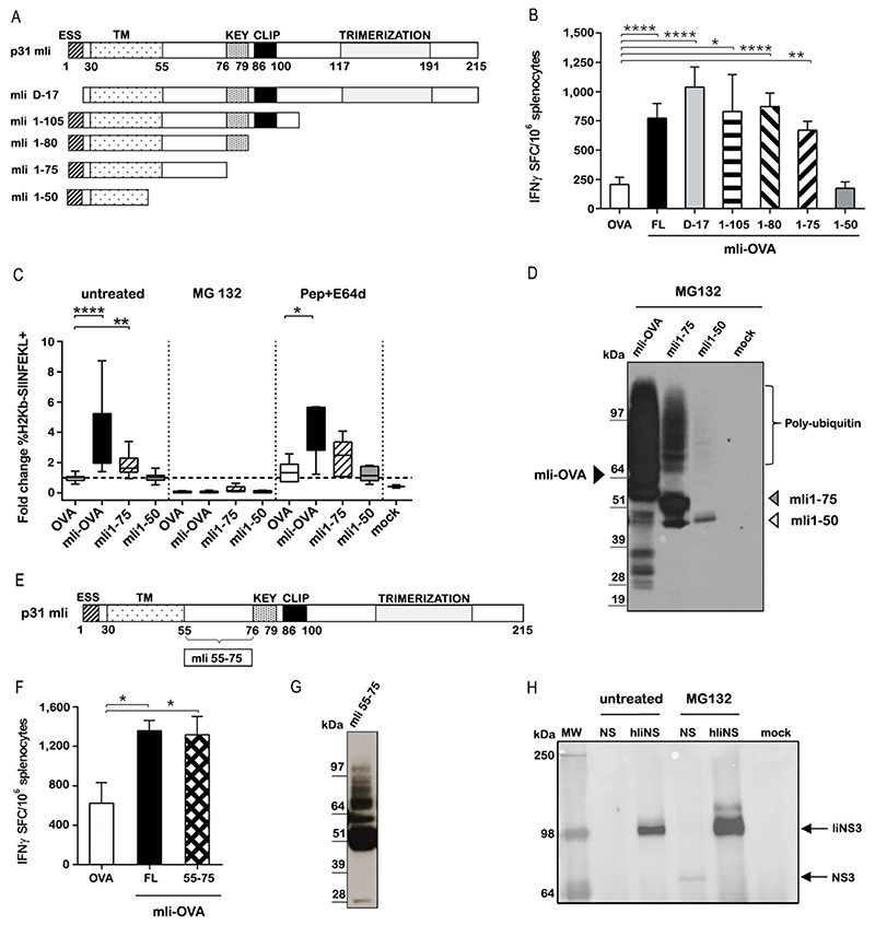
Figure 8. Ii modulates CD8+ T cell immune responses by targeting fused antigens for K48-specific ubiquitination and proteasome-mediated degradation.
(A) Schematic representation of full-length murine p31 Invariant chain (mIi) and deletion mutants. Functional domains are indicated: ESS, endolysosomal sorting signal; TM, transmembrane domain; KEY, key motif; CLIP, class II-associated Ii chain peptide; Trimerization, trimerization domain. (B) C57BL/6 mice were vaccinated with 3x10^6 viral particles of Ad5 encoding OVA either alone or fused to the indicated mIi deletion mutants. T cell responses were evaluated 2 weeks later by IFN-γ ELISpot assay. Data are expressed as number of T cells producing IFN-γ per million splenocytes (Ordinary one-way ANOVA test for FL, D-17, 1-105,1-80, 1-75 mIi constructs versus OVA) The experiment were repeated four times. (C) The percentage of Ad5 infected CD11c+ BMDC cells expressing SIINFEKL peptide bound to H-2Kb MHC class I left untreated or treated with MG132 (proteasome inhibitor) or Pepstatin A/E64 (lysosomal proteases inhibitor) was evaluated. Results are expressed as fold difference relative to Ad5-OVA infected cells (Kruskal-Wallis test untreated OVA vs mIi-OVA; OVA vs mIi1-75; Pepstatin+E64d treatment OVA vs mIi-OVA) The experiments were repeated five times in duplicate. (D) HeLa cells transiently transfected with ubiquitin plasmid were infected with Ad5-mIi-OVA, mIi1-75 OVA and mIi1-50 OVA and treated with MG132. Cells extracts were immunoprecipitated with anti-Lys48 antibody and analysed by WB with an anti-HA antibody detecting OVA. (E) Schematic representation of mIi and 55-75 region. (F) Immunogenicity in C57BL/6 mice of Ad5-mIi55-75 was evaluated after 2 weeks later by IFN-γ ELISpot assay (Ordinary one-way ANOVA mIi OVA versus OVA; mIi55-75 OVA versus OVA). The experiments were performed three times. (G) HeLa cells were transfected with ubiquitin plasmid, infected with Ad5 -mIi55-75 and treated with MG132. The cells lysate was immunoprecipitated using an anti-Lys48 antibody and tested by WB using anti HA antibody detecting OVA. The experiments were performed two times. (H) HeLa transiently transfected with ubiquitin plasmid were infected with ChAd3-NSmut and with ChAd3-hIiNSmut and treated or not with MG132. Cells extracts were immunoprecipitated with anti-Lys48 antibody and analysed by WB with an anti-NS3 Ab detecting NS3 protein. Mock is negative control for uninfected cells. The experiments were performed three times. *P ≤ 0.05; **P ≤ 0.01; ****P ≤ 0.0001

