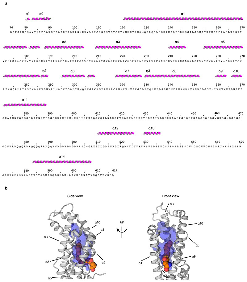
Extended Data Figure 7. Additional structural information on Dlpcore in complex with hWnt7a peptide.
a) Sequence of Dlpcore construct from the complexed structure annotated with secondary structure elements. To facilitate comparison between the complexed and apo structure, secondary structure nomenclature of the complex reflects that of the previously published apo structure (PDB ID: 3odn). α indicates alpha-helices, η indicates 310-helices. b) Side and front view of the lipid binding cavity of Dlpcore in complex with the Wnt palmitoleoylated peptide, showing the cavity extension beyond the end of the Wnt peptide acyl chain. This additional space likely accommodates the bodipy moiety of bodipy-palmitate for the assays presented in Fig. 4a. The internal volume of the cavity is coloured in blue, with the palmitoleoylated serine from the Wnt peptide represented as spheres in atomic colouring (C: orange, N: blue, O: red).

