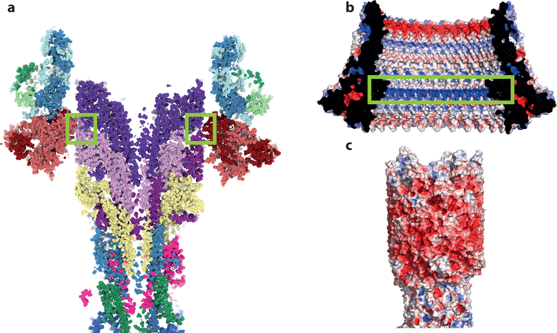
Extended Data Fig. 6. Structure of the LP-rod bearing.

a, Slab through the LP-rod demonstrating the thin seal around the rod. Green boxes highlight the constriction point formed by residues 48–82 of FlgI. b, Electrostatic potential mapped onto the surface of the LP-ring (view from inside the ring) with the positively charged band at the seal point highlighted in a green box). c, Electrostatic potential mapped onto the surface of the rod. Electrostatics analysed using APBS within Pymol.
