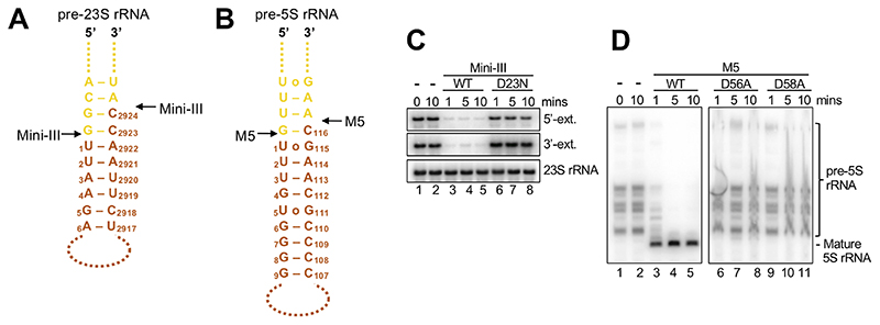
Figure 1. rRNA precursor substrates for Mini-III and M5.
(A) Schematic outline of pre-23S rRNA substrate helix highlighting the nts of the 3’-and 5’-extensions in yellow, and mature 23S rRNA nts in orange. Only overhang nts built in the cryoEM models are shown. Mini-III cleavage sites are indicated with black arrows.
(B) As for (A), but for pre-5S rRNA/M5.
(C) Northern blot showing removal of 5’-and 3’-extensions from 50S(pre-23S) particles by wild-type Mini-III (Mini-IIIWT) (lane 3-5), and lack thereof by Mini-IIID23N mutant protein (lane 6-8) at different time points. Lanes 1-2 show pre-23S rRNA at the first and last time point in the absence of Mini-III protein.
(D) As for (C), but for M5WT and M5D56A/M5D58A mutant proteins with 50S(pre-5S) particles.

