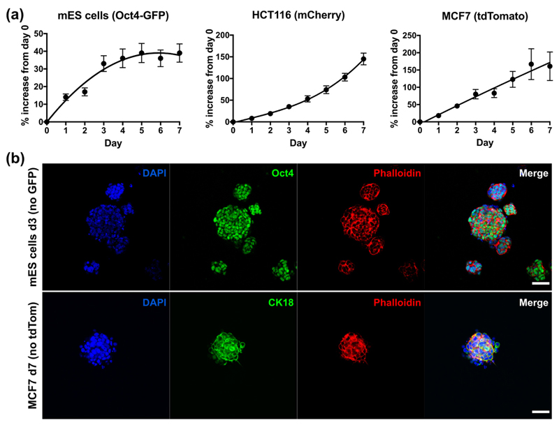
Fig. 3.
Methods for assaying cell growth and pluripotency in the 6 mg/mL peptide gels. (a) Real-time measurements of % increase in signal relative to day 0, from fluorescently-tagged cell lines encapsulated in matrix-free gels (seeding density 5 × 10⁵ cells/mL). Graphs show mean ± standard deviation for n = 3 independent experiments. Trendlines are intended as a visual guide only. (b) End-point immunostaining and microscopy of unmodified mES (Oct4 stain) and MCF7 (CK18 stain) cell lines seeded within the matrix-free peptide gels. Scale bar 50 μm.

