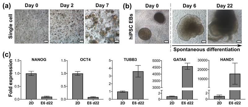
Fig. 4.
hiPSC growth and differentiation seeded in matrix-free 6 mg/mL peptide gels. (a) hiPSC seeded as a single cell suspension at 1 × 10⁶ cells/mL, (b) embryoid bodies (EBs) formed of 2000 cells per EB, seeded at 8–10 EBs/gel and maintained in E6 medium. Scale bar 100 μm. (c) qPCR results for EBs maintained to day 22 in E6. Fold expression is shown relative to hiPSC grown in 2D on vitronectin to day 3, normalised to RPLPO. OCT4 and NANOG are used as markers of pluripotency, whilst TUBB3, GATA6 and HAND1 are used as markers of ectoderm, endoderm and mesoderm respectively. Graphs show mean ± SEM for n = 2 independent experiments.

