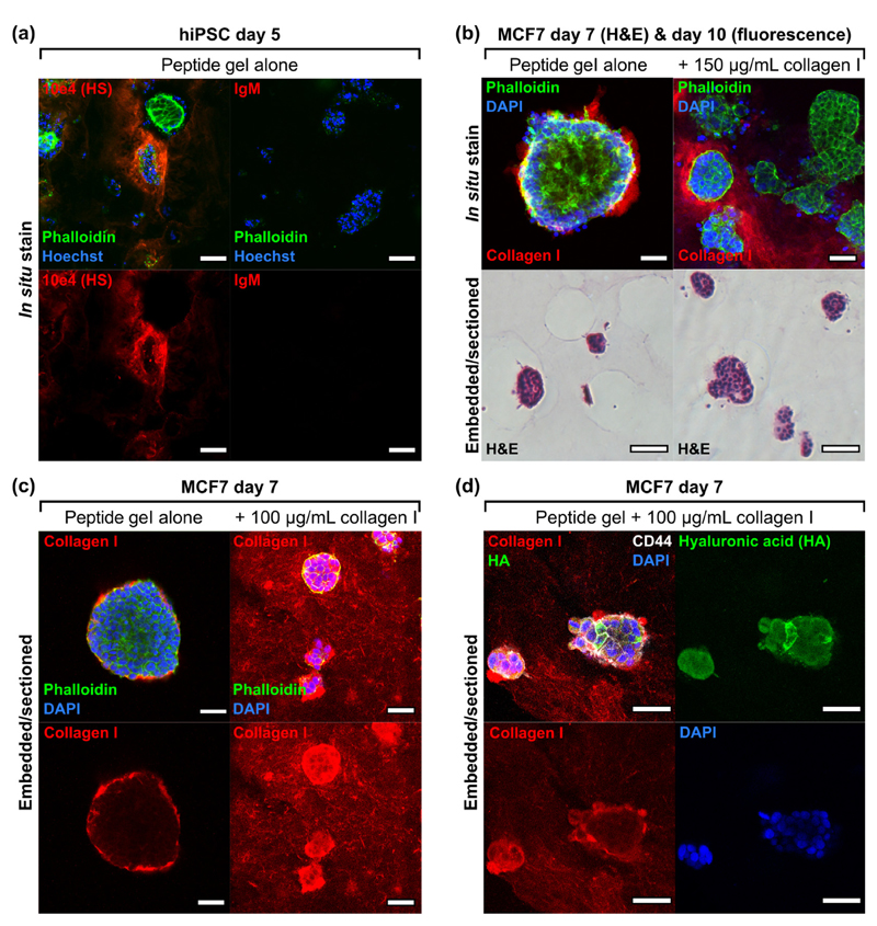
Fig. 6.
Exogeneous and endogenous matrix may be distinguished using immunostaining in 6 mg/mL peptide gels. (a) Heparan sulphate (HS) deposition by human induced pluripotent stem cells (1 × 10⁶ cells/mL), (b) collagen I localisation on culture with MCF7 in unmodified peptide gel or with collagen I modification, with corresponding H&E (5 × 10⁵ cells/mL), (c) improved collagen I localisation on embedding and sectioning (seeding density reduced to 1 × 10⁵ cells/mL for 100 μg/mL collagen to avoid overconfluence at day 7 in this condition), (d) co-stain for exogenous collagen I and endogenous HA (biotinylated hyaluronic acid binding protein, bHABP, detected using TRITC-streptavidin) in peptide gel modified with collagen I (1 × 10⁵ cells/mL). Negative control images can be found in Supplementary Fig. 4. Scale bar 100 μm for fluorescence images in panel (b), otherwise all scale bars are 50 μm.

